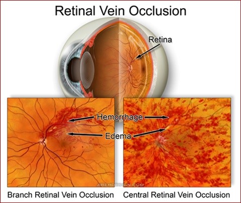
The retina is the photographic film of the eye. It is covered in arteries carrying blood to the eye, and veins carrying blood away from the eye. There is one main central artery and vein, and multiple smaller arteries and veins coming from this central trunk. If one of these veins become blocked, then the backpressure in the system causes the fine retinal capillaries to break, resulting in haemorrhage and leakage of fluid (oedema). If this occurs in a tiny retinal vein supplying peripheral vision, then it may occur without you realising, and may be of little consequence. However, if the blockage occurs in one of the larger veins, or even the central retinal vein, then this will cause sudden and profound loss of vision.
What causes a blocked vein?
Blocked veins are caused by the same things that cause other cardiovascular problems, such as heart attacks and strokes. All of these things are caused by a combination of factors, including increasing age, poor diet, lack of exercise, smoking. It is also caused by things like high blood pressure and cholesterol. These factors cause gradual accumulating damage to the lining of blood vessels, and ultimately, cause a blood clot to form in the vein. This clot blocks the whole vein.
Although blood clots usually dissolve, the short period of time that the vein was blocked was enough to damage the retinal circulation significantly. This damage may take years to recover. As the retina heals some sight may return, but in others, the sight either does not recover, or gets progressively worse.
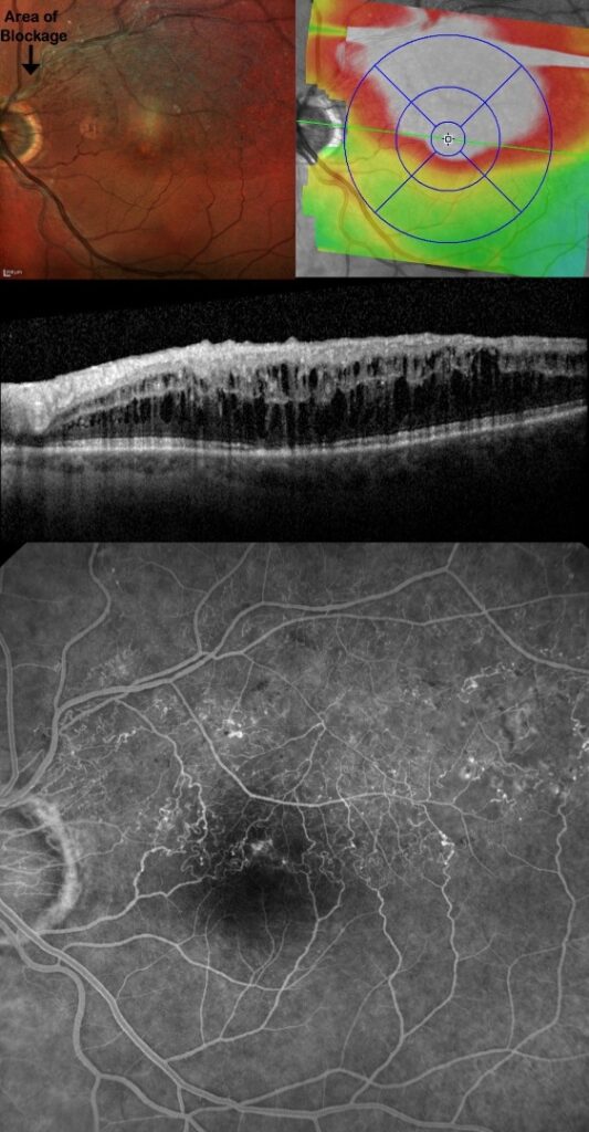
Patient with a blocked vein in the left eye. The colour photograph illustrates the swollen appearance of the superior half of the macula. The swelling is coloured in the overlay on the right. Beneath this is the OCT of the macula showing gross retinal oedema (swelling). A fundus fluorescein angiogram (bottom image) shows the damaged blood supply of this part of the retina.
What is the treatment of a vein occlusion?
Once reversible causes have been ruled out treatment can be started. Treatment comes in 2 forms, laser or injections into the eyes. Laser therapy is useful if you have a small branch vein occlusion. Gentile stimulating laser of the macula can trigger the retina to absorb the swelling and help the retina to dry out. Injections are now the mainstay of treatment for this condition. Injections can be of either a 4-6 month dissolvable implant of anti-inflammatory steroids, such as Ozurdex, or a series of monthly anti-VEGF injections, such as Avastin, Lucentis or Eylea.