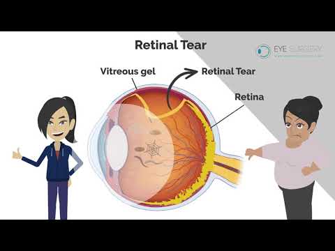
The retina is a thin sheet of photographic film that lines the inside of the eye. The cavity of the eye is filled with a transparent gel, known as the vitreous. When we are born this vitreous gel is adherent to the retina and fills the whole cavity of the eye. As we age the vitreous gel shrinks and dissolves. There comes a point, usually in midlife, when the gel has shrunk so much that it can no longer fill the whole cavity. Hence, it naturally separates from the retina lining the eye in a process known as a posterior vitreous detachment (PVD). This PVD occurs in everybody and is a normal part of aging. As a PVD develops you may experience some flashing of lights as the gel pulls on the retina. Eventually, when the PVD has completed you will have more floaters that usual. These floaters are the back surface of the gel, which used to be attached to the retina, but is now moving in front of the retina.
In a small percentage of people, as the gel peels away from the retina, if the gel is too adherent at any one point, the retina can tear. A retinal tear is very serious, as the retina is largely glued to wall of the eye by vacuum. A tear to the retina will break the vacuum. This allows the retina to slowly peel away from the back of the eye. This is known as a detached retina. This is an emergency.
What are the symptoms of a retinal tear?
When the retina tears it will bleed. Blood would appear in the eye as multiple dense floaters. These are not like the normal occasional extra floater that comes and goes. Floaters caused by a retinal tear are many and dense. It is described as a ‘shower’ of floaters. If you have a sudden shower of floaters, get seen within 24 hours to rule out a retinal tear. If the tear is caught early then it can simply be sealed by laser in clinic. If it is not caught in time, and the retina has already started to detach, it will require an operation to fix.
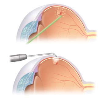
A tear to the retina, if cught early enough can be simply lasered in clinic to seal it and prevent a retinal detachment.
What are the symptoms of a retinal detachment?
If the retinal tear has not been detected in time, and the retina has started to detach, then you would see a slowly enlarging shadow in your peripheral vision. This can be described as a hazy curtain being slowly drawn across the eye. The vision may remain sharp for a little while. However, if the detachment spreads to reach the central vision, which may occur in as little as 1 day, then the vision will be significantly impaired.
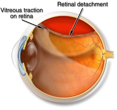
Once the retinal detachment has reached the central vision, there will be some permanent damage to the sight. If the retinal detachment is repaired much of the sight can be restored, but it will never be as good as it was again.
The emphasis is clearly therefore of catching tears or detachments early to prevent loss of sight. The longer the retina remains detached, the worse the visual prognosis will be. Most units try and repair retinal detachments within 24 hours. If the central vision has become involved then the opportunity to keep perfect vision is lost, and whilst still urgent, the retina can be repaired within a couple of days.

A photograph of a detached retina. The sheet of undulating retina that has detached can be seen superiorly. The detachment in this case extends right through the central macula, with some retinal wrinkles visible beneath the fovea. Some blur and distortion will remain, despite retinal detachment repair because of this.
How is a retinal detachment treated?
The treatment for a detached retina is a vitrectomy operation. During a vitrectomy three tiny pinprick size (0.5mm) holes are made in the white of the eye (sclera). Through this the vitreous gel, that caused the detached retina, is removed. The fluid is drained from underneath the retina to recreate the vacuum. The retinal tear is sealed with laser or cryotherapy, and the eye is filled with a temporary internal gas bandage. The gas expands inside the eye, pushing the retina against the wall of the eye. The gas bubble remains there for at least a week, or until the retina has healed. The gas bubble slowly and naturally dissolves and disappears.
When the eye is full of gas there will be very little sight because the eye is not designed to look through gas. As the gas dissolves, you will become aware of a line appearing in the top of your vision. This line will wobble and move around. This line is the bottom of the gas bubble and your eye is effectively a human spirit level. Each day the line will get lower and lower and begin to round up. Above the line the vision will start becoming clearer. The line is the most annoying as it passes through the centre of the vision about 1-2 weeks after surgery. The bubble will eventually become circular and break up and disappear.
How successful is retinal detachment surgery?
Retinal detachment surgery is successful 85% of the time after a single procedure. This means that 15% of detachments will recur. The retina may require several operations to repair and long acting gas bandages or silicone oil may be used in extreme cases. The vision cannot be guaranteed. Hence the importance of detecting tears early and treating them, rather than allowing the retina to detach.