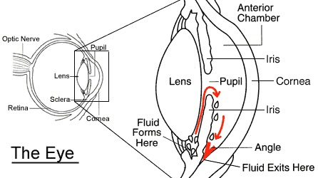
The eye is a fluid filled structure. Just like a football or tyre, it can be under filled and soft, or over filled and hard. The fluid that fills the eye is known as aqueous fluid. This is made at the back of the eye by the ciliary body. This aqueous fluid circulates through the pupil into the anterior chamber, and drains out of the eye near the root of the iris, where it meets the cornea. This is known as the “angle” or “trabecular mesh.”
If too much fluid is produced, or the drainage system is blocked, then the eye pressure will rise. This is known as ocular hypertension. The normal eye pressure is measured in millimetres of mercury (mmHg). The normal pressure is 10 to 21mmHg. Anyone with a pressure over 21mmHg has ‘ocular hypertension’. If it is only a little over 21mmHg then this is unlikely to be harmful to the eye and can often be safely monitored without treatment, although this depends on a number of variables. The higher the pressure within the eye the more likely it will cause pressure related damage to the eye. If there is any evidence of damage to the eye from raised pressure, the condition is termed Glaucoma rather than ocular hypertension. Glaucoma requires treatment. There are various treatments for ocular hypertension and glaucoma. They vary from eye drops, to laser of the iris (iridotomy) or trabecular mesh (trabeculoplasty), or to surgery (i-stent or trabeculectomy).
Eye drops work by either reducing the production of aqueous fluid by the ciliary body, or increasing the drainage of fluid from the eye. Laser of the iris on the other hand physically makes holes in the drainage pathways within the eye, allowing aqueous fluid to escape more quickly, thereby lowering eye pressure. Laser for ocular hypertension or glaucoma is either known as an iridotomy (hole in the peripheral iris) or trabeculoplasty (hole in the trabecular meshwork).