Eye Surgery Ltd is the web site of
Mr Steven Harsum
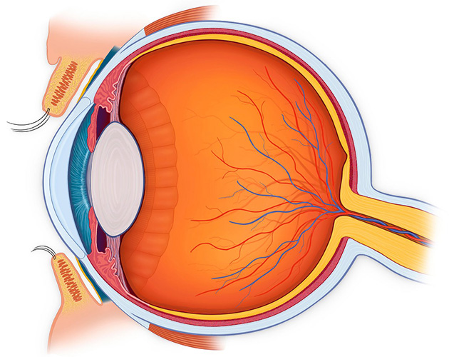
Click the picture to learn about it

Optic-Nerve
This connects the photographic film of the eye (retina) to the brain. It has over 1 million nerve fibres in each optic nerve.
Problems with the optic nerve typically cause loss of fine vision, loss of colour vision, and loss of field of vision. The nerve may be damaged by eye pressure (glaucoma), inflammation and swelling (optic neuropathy & papillitis), tumours around the nerve, or raised brain pressure (papilloedema).
Sclera
The wall of the eye is a tough white layer known as the Sclera. This can become red and inflamed. Superficial inflammation is known as episcleritis and deeper inflammation is known as scleritis. These may be associated with generalised inflammation within the body, such as rheumatoid arthritis, or sarcoidosis.
The length of the eyeball from front to back determines whether you are long sighted (a short eye) or short sighted (a long eye).
Choroid
This is a vascular layer beneath the retina that supplies the retina with oxygen. Problems with the choroid include haemorrhage, and abnormal blood vessel gmap-rowth, that leads to a type of wet macular degeneration .
Choroid
This is a vascular layer beneath the retina that supplies the retina with oxygen. Problems with the choroid include haemorrhage, and abnormal blood vessel gmap-rowth, that leads to a type of wet macular degeneration .
Retina
This is the thin delicate photographic film of the eye that covers the inside of the eye. The finest vision is achieved at the very central focal point of the retina, known as the macula.
Problems with the retina present with distortion, blur, or shadows in the vision. Torn or detached retinas are a surgical emergency. A torn retina may cause a ‘shower’ of floaters (retinal tear), a curtain-like peripheral shadow (macula sparing retinal detachment ), or loss of vision (macula involving retinal detachment). Age related macular degeneration (AMD) is where the retina becomes thinned (Dry AMD) or swollen (wet AMD). AMD and diabetic retinopathy, are the commonest causes of blindness in the UK. Other causes of retinal damage are blocked veins or arteries.
Vitreous
This is the clear gel that fills the back of the eye. When you are born it completely fills this space and is attached to the photographic lining of the eye known as the retina. As you age the gel dissolves and shrinks.
Problems with the gel are related to the accumulation of debris within the gel (floaters), and the natural shrinkage and subsequent separation of the gel from the back of the eye known as vitreous detachment.This may cause a sudden increase in floaters or may stretch (macular traction) or tear (retinal or macula hole) or scar (epiretinal membrane) the retina within the eye. It may also tear a blood vessel and cause vitreous haemorrhage
Lens
Just as you have a lens in your glasses to focus there is a lens in the eye to focus. When you are young this changes shape to bring things into focus. As you age it stops changing shape and reading glasses are subsequently required (presbyopia).
Problems occur when the lens loses its clarity and becomes opaque. This causes blurring that can not be corrected with glasses. This is known as a cataract. Cataract surgery is the commonest operation in the country. After cataract surgery, the capsular bag that supports the lens can become cloudy. This posterior capsule opacification blurs the vision and requires a minor YAG laser procedure in clinic.
Zonules
The zonules are elastic fibres that anchor the lens of the eye in place. They act rather like springs around the edge of a trampoline, holding the lens centrally in the eye. If the zonules are damaged by a medical condition called pseudoexfoliation, surgery, or trauma, then the lens will no longer be stable and can move out of position.
Ciliary Body
The Ciliary body is the structure in the eye that produces fluid. Fluid is made here, behind the iris, flows through the pupil into the anterior chamber, and drains out of the eye at the trabecular mesh at the root of the iris. This fluid controls the https://eyesurgeryltd.co.uk/2020/12/28/high-pressure-ocular-hypertension/pressure of the eye .
Ciliary Body
The Ciliary body is the structure in the eye that produces fluid. Fluid is made here, behind the iris, flows through the pupil into the anterior chamber, and drains out of the eye at the trabecular mesh at the root of the iris. This fluid controls the pressure of the eye .
iris
The iris is the part of the eye that gives it colour. The centre of the iris is the pupil, where light enters the eye. As the iris acts as a shutter, controlling the size of the pupil and the amount of light that enters the eye.
Inflammation of the iris is known as anterior uveitis or iritis. Iritis causes a red and painful eye with blurred vision and photophobia. At the root of the iris in the anterior chamber is the trabecula mesh. The trabecular mesh is where fluid drains from the eye and enters the blood circulation.
Anterior Chamber
This is the fluid filled front chamber of the eye. The fluid, known as aqueous, is produced behind the iris by the ciliary body, flows through the pupil, and drains out near the root of the iris, at the trabecular mesh.
Problems with drainage of fluid from the eye causes the pressure in the eye to rise. High eye pressure is termed ocular hypertension. As the pressure rises this may damage the sight, known as Glaucoma. Rarely, the pressure in the eye can rise within hours, causing immediate loss of vision with a painful red eye. This is known as angle closure glaucoma and must be treated urgently.
Cornea
The cornea is the clear window covering the surface of the eye. The shape of this contributes towards the sharpness of your vision. If it is irregular (rugby ball shaped rather than football shaped), this is known as astigmatism. This astigmatism can be corrected with glasses, contact lenses, or refractive laser (LASIK & LASEK) to the cornea.
The clarity of this clear window can be disrupted by degeneration or dystrophy.
Corneal problems cause watering and blurring of vision. Infections or scratches cause intense pain and photophobia. Scratches are invisible whereas infections cause a white patch to appear on the cornea. Corneal infections, known as corneal ulcers, are an urgent sight threatening condition that usually occurs in contact lens wearers.
Conjunctiva
The conjunctiva is a very thin transparent layer covering the sclera and the inside of the eyelids. The conjunctiva is mobile and the blood vessels of the conjunctiva can easily bleed causing a subconjunctival haemorrhage. Infections of the conjunctiva are known as conjunctivitis. Swelling and ballooning of this layer is called chemosis. Thickening of the conjunctiva is called pingueculum and conjunctiva that abnormally gmap-rows over the cornea is called a ptygerium.
Extraocular Muscles
There are 6 muscles surrounding the eye that move the eye around. Disorders of the wiring or higher control of these muscles causes a squint, where the eyes stop working as a pair, and they become misaligned. This may cause binocular double vision.
Extraocular Muscles
There are 6 muscles surrounding the eye that move the eye around. Disorders of the wiring or higher control of these muscles causes a squint, where the eyes stop working as a pair, and they become misaligned. This may cause binocular double vision.
Lids
The eyelids are essential for keeping the eye moist and lubricated. They contain large oil glands known as meibomian glands. Meibomian oil mixes with tears to form a protective tear film. Lack of tears or oil causes dry or watery eye. Tear ducts in the upper and lower lids drain excess tears from the eyes to the back of the nose and throat. Blockage of these tear ducts can also cause watering
The oil glands within the eyelid margins can become inflamed or blocked (blepharitis) causing dry eyes. They can become infected causing a tender lid lump (Stye). The upper lid may droop (ptosis) and obscure the vision or become elevated (retracted) giving a staring appearance. The lids can lose their elasticity, allowing them to roll inward (entropion) or outward (ectropion). This causes the eye to become red and sore.
What are Cataracts
What does cataract surgery involve
Risks of cataract surgery
Retinal tear
Lens implants
Glasses after surgery
What is Astigmatism
Bilateral surgery
Do I need surgery
Epiretinal Membranes
- About Me
Eye Surgery Ltd is the web site of Mr Steven Harsum, a substantive consultant ophthalmologist specializing in cataract and retinal surgery, based in South London. This web page is designed to be informative and educational.
Click the eye structure about to learn more about its different parts and the medical conditions related to each part.
See animations and videos explaining cataract and retinal surgery here or on my YouTube channel .
Test your vision at my website www.testmyvision.co.uk. Here you can test your distance and reading vision, measure your distortion, and draw your floaters.


Our Locations
Ashtead Hospital, The Warren, Ashtead, Surrey, KT21 2SB
St Anthony's 801 London Road, Cheam, Sutton, Surrey, SM3 9DW
Epsom General Hospital, Eye Unit, Woodcote Wing, Dorking Road, Epsom KT18 7EG
St Helier Hospital, Wrythe Lane, Sutton, Carshalton SM5 1AA Ophthalmology Outpatients Department, Ground Floor B-block Ophthalmology Day Surgery, 4th Floor B-block
Nuffield Health Parkside Hospital, 53 Parkside, Wimbledon, London, SW19 5NX
140 London Rd, Kingston upon Thames KT2 6QL
Reviews
I see colours now that I’d forgotten existed My first contact with Mr Harsum was in the Spring of 2021. When I knew in July this year that I needed to have a cataract operation I telephoned his Secretary and was fixed up immediately at Ashtead Hospital. She is very efficient and helpful. After my Consultation with Mr Harsum when he explained what he would do in the two operations my mind was completely at rest. He is very positive and knowledgeable and dates were fixed. I cannot praise him enough as I have excellent vision now and have no need for glasses at all. Colours are brilliant and I have real depth of vision. The best since I was 11 !! Thank you so much for your expertise and care.
Mr Harsum performed surgery on my eyelid and surrounding area. Mr Harsum performed surgery on my eyelid and close area, he did this skillfully and my eye looks normal again. His manner and all the staff at Ashford Hospital were kind, calm, and caring and put me at ease straight away, couldn't be happier!
The Perfect Experience We saw Mr Steven Harsum at Ashstead recently after my wife was unequivocally diagnosed with Wet Macular Degeneration by a senior private consultant elsewhere. This (faulty, as it turns out) diagnosis was seemingly catastrophic for my wife, condemning her to a lifetime of monthly intra ocular injections and inevitably failing eyesight at the age of 57. In a wonderful Doc Martin moment, after examination and scans, Mr Harsum re-diagnosed my wife's issue as far less concerning CSR, not Wet MD. In fact, I suspect he already knew this even prior to our arrival, having requested my wife's historical opticians scans. In sum. I cannot recommend him highly enough - massively competent, pleasant and diligent. Mr Harsum's competence appears to have even rubbed off on his secretary - who expressed some suprise on the first contact call that my wife might have Wet AMD at her age. 10 stars, if they were available.
I thoroughly recommend Mr Steven Harsum… I cannot recommend Mr Steven Harsum enough, excellent treatment and a very nice man. I am absolutely delighted with my 'new' eyes. His secretary Katherine Gillan was fantastic too, finding me appointments and keeping in touch through the Covid 19 lockdown, which happened just after I had my first eye done. If you need lens replacement, don't go to anyone else!!!!
